Electrophoresis
Electrophoresis is carried out with equipment consisting of a negative charge at one end and a positive charge at the other, called an electrophoresis system. When charged molecules are inserted into this environment, the negatively charged molecules will go to one end and the positively charged molecules to the other end. For example, when analyzing proteins in a gel, in these machines, the whole protein is taken to analyze its size. In this way, the shorter proteins will migrate to the poles more quickly and will be reflected at the bottom of the gel. The longer ones, on the other hand, will remain at the top.
When a mixture of ionized molecules with a net charge are placed in an electric field, they experience a force of attraction towards the pole with the opposite charge. After a certain time has elapsed, the positively charged molecules will move towards the cathode (negative pole) and the positively charged molecules will move towards the anode (positive pole).
 (1)")
Types of Electrophoresis a Laboratory May Need

Horizontales
Horizontal gel electrophoresis uses the basic theory for the separation of DNA, RNA or protein molecules according to their respective molecular size and charge. In this technique, the gel is present in a horizontal orientation and is immersed in a buffer that is continuous. The agarose gel is used to separate the gel box into two compartments. One end of the gel box contains an anode, while the other end contains a cathode. When a current is applied, the buffer used in this technique allows the creation of a charge gradient. When the charge is applied, the gel tends to heat up.
The buffer also functions as a coolant, which maintains the temperature at optimal levels. Recirculation of the running buffer prevents the formation of a pH gradient. A batch buffer system cannot be used in horizontal gel electrophoresis since the two compartments of the gel system are connected to the running buffer.
Verticales
The vertical gel electrophoresis technique works according to the primary theory of gel electrophoresis, but is considered to be more complex than the horizontal gel electrophoresis method.
This technique uses a discontinuous buffer. A cathode is located in the upper chamber, and the anode is located in the lower chamber. The electrodes present in each compartment provide the required electric field. A thin layer of gel is poured between the two mounted glass plates. Thus, the upper part of the gel is immersed in the upper chamber, and the lower part of the gel is immersed in the lower chamber. Once the current is applied, a small portion of the buffer moves into the bottom chamber from the top chamber through the gel.

In Kalstein you can find the ideal electrophoresis for your laboratory

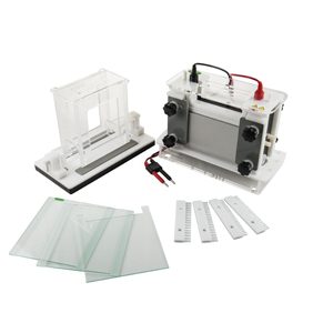
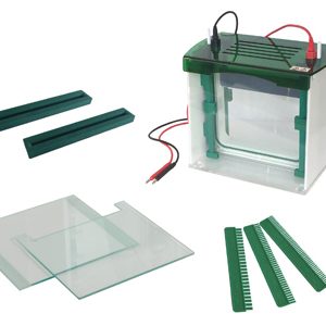
Mini-Protean Vertical Electrophoresis Cell YR03427
Vertical gel electrophoresis is a more complex set-up compared to horizontal gel system. Researchers normally...

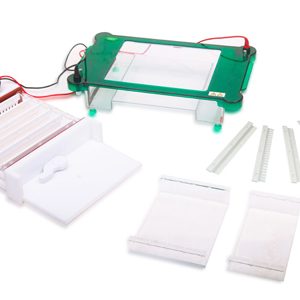
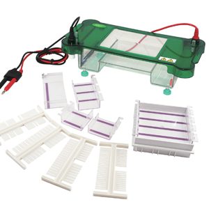
Rapid SSR Vertical Electrophoresis Tank YR03431
Vertical gel electrophoresis is a more complex set-up compared to horizontal gel system. Researchers normally...

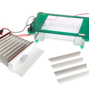
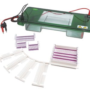
Horizontal Electrophoresis YR03421
YR horizontal gel electrophoresis units have been designed by scientists with the laboratory environment ...

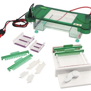
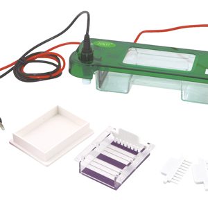
Horizontal Electrophoresis YR03420
YR horizontal gel electrophoresis units have been designed by scientists with the laboratory environment ...
Our Best Selling Electrophoresis
- The installation of the glass plate in just 15 seconds, and does not need the knob, fast and convenient.
- With the gel making device of normal position,don’t should to moving the glass plate from the process of making gel to electrophoresis.
- With the Precast gel inner core, do not affecting the operation of gel making while the electrophoresis.
- Safety cover open button design, convenient opening the cover.
- Abundant buffer not only guarantees cooling effects, but also keeps the pH Value stable during the whole experiment process.
- Auto-switch-off when the lid is removed.
| Model | YR03430 |
| Glass plate size(W×L) | 216×110 mm |
| Gel Size(W×L) | 186×95 mm |
| Gel thickness | 1.0(mm) |
| Gel amount | 1~2(pieces) |
| Sample volume | (1.0mm)25、40、52wells |
| Buffer Volume | ~1800(ml) |
| Dimension(L×W×H) | 280×140×135(mm) |
| Net weight | 3.2(kg) |

Analysis of the best Electrophoresis for your laboratory

What is electrophoresis?
Electrophoresis is an analytical method in which a controlled electric current is used in order to separate biomolecules according to their size to electric charge ratio, using a...

Electrophoresis Power Source
Electrophoresis is a basic method in the field of molecular biology for the analysis (separation, purification, preparation) of nucleic acids and proteins....

What is two-dimensional electrophoresis?
Two-dimensional electrophoresis is a special type of electrophoresis, considered a high-resolution technique that ...

Electrophoresis and Gel Documentation Systems
Gel electrophoresis is a widely used technique in life science laboratories to separate macromolecules such...
Electrophoresis models catalog on offer
Guides to Becoming an Electrophoresis Expert
Why is electrophoresis necessary in the laboratory?
Electrophoresis is an essential laboratory technique in biological studies, and its use is spreading to other types of fields ...
How does electrophoresis work molecularly?
Functionally electrophoresis is a technique that consists of proportional migration of molecules through a gel or other ...
Using Electrophoresis to Detect Genetic Diseases
In humans, the genetic link of certain diseases between sex-linked hemophilia and color blindness was first observed...
Horizontal and vertical electrophoresis: Differences
Gel electrophoresis is a laboratory technique used in genetics to separate mixtures containing DNA, RNA, and other proteins according to their respective molecular size and charge. The DNA, RNA, or proteins that must be separated in this method...
")
Frequently Asked Questions about Electrophoresis
How to know the prices of Electrophoresis?
To know the price of Electrophoresis we invite you to send us an email with your request through the contact form.
What are the delivery times of the Electrophoresis?
- If the equipment of your interest is in stock or if it must be manufactured.
- The type of freight you have chosen, this may be; air or sea.
How to make a purchase of Electrophoresis?
- By email: [email protected]
- By telephone: +33 (0) 1 78 95 87 02
- E.commerce: Via Kalstein's official website in your country.
How does the warranty work?
Can I request a quote online?
Of course, you can request a quote for the Kalstein team of your interest, directly from our official website. Once you have identified your preferred model, click HERE