In humans, the genetic link of certain diseases between sex-linked hemophilia and color blindness was first observed. However, the application of the principles of genetic linkage to diagnosis was limited until the richness of the genetic markers present in DNA could be easily detected using the DNA molecule itself. Two main types of DNA variation can be detected between different individuals or between a pair of homologous chromosomes of the same individual:
- Single nucleotide substitutions may substitute a base pair (pb) for a different pb.
- There may also be a difference in the number of bp. This difference may involve the deletion (or insertion) of a single bp or may be more extensive, involving hundreds or thousands of bp.
The size of the human genome (about 3 billion bp) and the frequency of variants/deletions in the sequence suggest that the total number of common variants should be in the tens of millions. Theoretically, markers linked to any disease gene can be analyzed by DNA typing.
However, as disease genes are localized by linked markers, this linking approach to genetic diagnosis is rapidly being overtaken by direct DNA typing of the disease’s own mutations. Direct diagnosis of mutations plays an increasingly important role in genetic diagnosis as more disease genes are being isolated and their mutations discovered.
Some typical genetic markers used in diagnosis
Restriction fragment length polymorphisms (RFLPs) were the first markers described to detect a variation in the structure of a DNA molecule. The length of a restriction fragment, generated by cutting the DNA with a given restriction enzyme (ER), will vary if:
- The enzyme recognition sequence has a nucleotide substitution that prevents cleavage.
- The number of bases between two cut sites by ER varies due to insertion/deletion.
Traditionally, RFLPs have been detected by hybridizing DNA probes, radiolabeled with phosphorus 32, to the Southern blots of restriction fragments separated by agarose gel electrophoresis. One of the first applications of RFLP markers in inherited diseases was for the prenatal diagnosis of sickle cell disease. Other markers of interest are:
- Minisatellites: consist of a 17-35 pb hypervariable “center” sequence that is present as a variable number of tandem repeats (VNTR), resulting in a highly polymorphic or hypervariable region (HVR). An HVR near the a-globin gene has been useful in the diagnosis of polycystic kidney disease in adults.
- Microsatellites: consists of one GT repetition on one strand and one CA repetition on the complementary strand. These short stretches of microsatellites, about 10-20 repeat units, vary by a multiple of 2 bp between the members of homologous chromosomes. This has been found within the dystrophin gene that is mutated in both Duchenne and Becker muscular dystrophy.
Detection and visualization of genetic markers
The detection of genetic markers for DNA typing poses two problems. In the first, it is necessary to visualize the region of particular interest of between the three billion bp of human DNA. And, second, it is necessary to be able to discriminate between slightly different variants of the same DNA fragment.
Visualization has traditionally been based on the exceptional affinity of a single-stranded DNA probe molecule for its complementary strand. DNA probes, radiolabeled with phosphorus 32, have been hybridized with immobilized single-stranded DNA in nitrocellulose or nylon membranes, with the bands corresponding to the hybridization regions detected by autoradiography.
An alternative approach uses PCR to amplify specific fragments that lie between a pair of primers on opposite and complementary strands. The product can be directly visualized followed by electrophoresis in an agarose gel containing ethidium bromide (EtBr). When irradiated in ultraviolet light, the EtBr dye that is bound to the DNA will produce a yellow fluorescence.
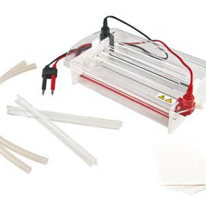
Why choose a Kalstein electrophoresis cell?
These cells are suitable to obtain the patterns of bright lines typical of the separation of DNA fragments obtained, since gel electrophoresis is an important technique to separate a large variety of molecules in genetics, and that offered by the manufacturer Kalstein, is suitable for different studies, such as the diagnosis of genetic diseases. This equipment is characterized by:
- Vertical or horizontal design, the latter being suitable for DNA and RNA analysis.
- Allows sufficient buffer solution to control the pH and cool the system.
- It has an auto off function when the lid is lifted.
The prices of these devices are really competitive and you can review more information such as purchase, quote or other technical details at HERE