Cardiovascular diseases are a frequent cause of death worldwide, including the pathogenesis of atherosclerosis (a specific type of arteriosclerosis). One of the phenomena related to the disease is the retention and subendothelial oxidation of cholesterol-rich lipoproteins. This process is associated with the onset of atherosclerotic plaque that is deposited in the carotid arteries reducing blood flow to the brain. Different types of cells are involved in arteriosclerotic disease. These include monocyte/macrophage endothelial cells, lymphocytes, smooth muscle cells, and platelets.
Arteriosclerosis can be one of the causes of myocardial ischemia, it occurs when there is complete or partial obstruction of the coronary artery by an accumulation of platelets; in the electrocardiogram it is expressed with a negative and symmetric T wave. However, other conditions can cause negativization of T waves.
The development and evolution of arteriosclerosis is accompanied by the presence of proaggregating, prothrombotic, mitogenic and proinflammatory effects that contribute to the formation of plaque in any artery of the body (of the heart, brain, arms, legs, pelvis and kidneys). This injury brings with it multiple conditions including coronary disease and carotid artery disease.
How is artery disease diagnosed?
Diagnosis is based on clinical physical evaluation by the cardiology specialist and is confirmed by angiography, ultrasonography, electrocardiography, imaging studies, and blood tests.
Electrocardiogram as diagnostic examination of arteriosclerosis
One of the most commonly used clinical studies to diagnose heart disease is the electrocardiogram, especially those related to heart rhythm and blood flow in the arteries. It is a basic, noninvasive scanning technique that is relatively straightforward and safe to use when interpreted correctly. There are several studies that try to define the relationship between electrocardiographic alterations and arteriosclerosis. Although to detect this abnormality the electrocardiogram is not a definitive study, the symptomatological context of the patient should be evaluated, if necessary with the help of complementary analyzes. An abnormal result may not necessarily be associated with heart disease.
Arteriosclerosis can be one of the causes of myocardial ischemia, it occurs when there is complete or partial obstruction of the coronary artery by an accumulation of platelets; in the electrocardiogram it is expressed with a negative and symmetric T wave. However, there are other situations that can cause negativization of T waves. If it becomes a lesion, the graph shows a ST segment elevation, which reflects a severe interruption of coronary flow to an area.
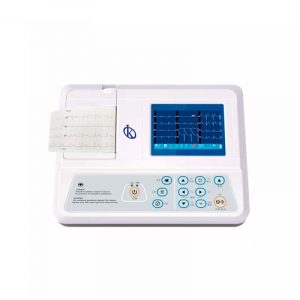
What is the Electrocardiograph?
The Electrocardiogram is the graphical representation of the electrical activity of the heart, in which differences in voltage are studied as a function of time (about 30 seconds). These changes result from depolarization and repolarization of the heart, which are caused by electric fields reaching the surface of the body where electrodes are placed.
The instrument associated with this technique is called the electrocardiograph. A team developed in 1903, by the Dutch doctor Willem Einthoven, and with the passage of time was evolving to the modern electrocardiograph. It works electronically and serves to detect possible faults in the heart. Recording of the heart’s electrical activity is done either in print (on strips of millimeter paper) or on the computer’s digital display (if modern). In both cases, it is observed in the form of a path that presents different wave deflections, which correspond to the path of the electrical impulses generated by the heart.
How is an electrocardiogram done?
To perform an electrocardiogram is a simple procedure, for this you need an electrocardiograph with the respective accessories for measurement, and proceed as follows:
- The patient lies on a stretcher horizontally, with the chest uncovered.
- The technician places 10 to 12 patches, spread across the chest and sometimes the arms and legs.
- Cardiac activity is displayed on the computer monitor or printed on paper.
At KALSTEIN we are MANUFACTURERS and we have at your disposal new electrocardiographic units. If you are interested in knowing our products, the PRICES for BUY or SALE visit our website HERE