Obtaining the histological material to study
Tissue processing is a very important part of any histology laboratory, and begins with obtaining the tissue under study. In the case of plant tissues, samples of the different organs that make up the body of the plant are taken directly, while human and animal tissues can be obtained in different ways through biopsies (excisional, incisional, endoscopic, colposcopic, etc. .) and by autopsy / autopsy.
Tissue fixation
The samples are usually fixed with liquid solutions called fixatives, which are used to keep the cellular and molecular structures unchanged during further processing and with an organization as similar as possible to how they were in the live sample.
Washes
The tissue should be washed to remove excess fixative (chemical). Excess fixative during the subsequent infiltration process, even in the microtomy, could affect the histological cuts, and therefore should be washed with distilled water.
Dehydration
Dehydration is performed using different alcohol solutions at increasing concentrations until reaching pure alcohol.
Clearing
In this step the alcohol is replaced by a substance that acts as an intermediary between the water and the paraffin that will be used later for infiltration. The commonly used substance is xylol although other organic solvents such as benzene can also be used. During this process the tissue loses color resulting in the term of clearance.
Infiltration
The sample is placed in histological paraffin in a liquid state, which is achieved by heating the paraffin above its melting point. Dehydration, clearance and infiltration can be done manually but nowadays they are done automatically on specific machines.
Inclusion
Its purpose is to provide the fabric with a solid support that makes it possible to make a very fine cut, so it is very important that the medium used for inclusion penetrates into the tissue. This is achieved by embedding the tissue with liquid substances that will subsequently polymerize (resins) or become consistent (waxes).
Microtomy
After inclusion or freezing, the tissues are cut, that is, sections are obtained. There are different cutting devices that allow sections of different thicknesses to be achieved: ultra-thin (of the order of nm), semi-thin (0.5 to 2 µm), thin (between 3 and 10 µm) and thick (larger than 10 µm).
Staining
The cells, by themselves, have no coloration. Therefore, in order to observe the tissue morphology, they must be “dyed.” There are many types of stains to differentiate the different structures or substances in the tissues.
The most commonly used staining is hematoxylin and eosin. Hematoxylin stains acidic substances or structures that contain them, such as the nucleus containing deoxyribonucleic acid (DNA) and eosin stains basic structures such as cytoplasm and other eosinophilic organelles in the cell.
Observation
Processed tissues are observed with microscopes. There are two basic types of microscopes: optical and electronic. The former offer great versatility in terms of ways of observing tissues, while the latter allow a high resolution power, being able to observe ultrastructural characteristics such as cell membranes or even molecular complexes.
The histological technique is widely used in laboratories and hospitals. It allows diagnosis of different diseases, such as to know if a tumor is malignant or benign, etc.
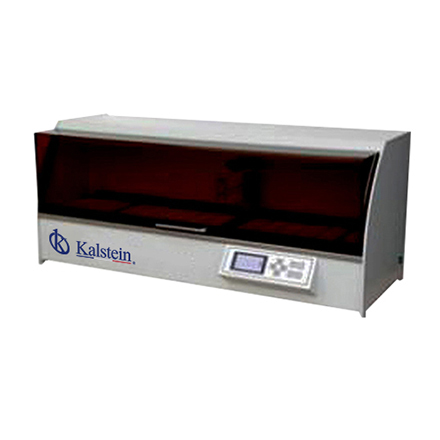
A tissue processor is an instrument that is used to analyze and process samples by fixing, staining, dehydrating or decalcifying them.
These devices have evolved slowly to be safer to use, handle larger sample numbers, process faster and produce better quality results. Most modern fluid transfer processors employ high temperatures, effective fluid circulation and incorporate vacuum / pressure cycles to improve processing and reduce processing times.
In Kalstein we have available tissue processors that are very useful in the histology laboratory. You can get more complete information about them through the following link HERE