The microtomes are cutting instruments for the preparation of preparations that are used in microscopy. To meet the high demands of such preparations, they allow extremely thin cuts. Normally modern microtomes allow cuts of a thickness of 0.1 to 100 µm. By way of comparison: Human hair has a thickness between 50 and 70 µm.
What is the history?
The history began with the start of light microscopes. In order to analyze objects, they must be fine enough for light to pass through. The first microtomes were initially simple blades (usually razors) with which cuts were made manually. As the demands on the preparations were increasing, it was necessary for the microtomes to develop. The first microtomes, as we know them today, were developed in 1770. With these you could fix the test and adjust the thickness of the cut by means of screws.
Nowadays
Today, mechanical microtomes are composed of a block, a sample holder and a technical equipment for the control of the advance. The quality of the preparations depends on the type of feed, the geometry of the blade and the declination (angle between the blade and the cutting direction). Additionally, the result can be influenced in the preparation of the sample (for example by freezing). In addition to mechanical microtomes, laser microtomes are increasingly used today, with which it is possible to prepare non-contact samples.
There are several types of microtomes (hand, balancing, rotating, sliding, cryostat, ultramicrotome, etc.). The rotation is the most used due to the advantages it provides: high precision and the possibility of producing very thin series sections, thanks to the demultiplication that produces the change of a movement of rotation to another of translation.
What kind of blades do microtomes have?
The blades can be made of three types of materials, which depend on the need that the laboratory needs to cover and the fineness of the cut required by the sections.
Steel blades
The steel blades are special for cutting soft tissue sections of animals and / or vegetables.
Steel blades can be used in histology, cork, wood and expanded polystyrene for light microscopy.
Glass blades
The glass blades are ideal for extracting very thin sections for light microscopy and electronics.
Diamond blades
Diamond blades for microtomes are generally used in industries, as these are used for fine cuts of hard materials such as bones, teeth and plant material such as hardwoods, as they are also ideal for light microscopy and electronics.
How does a rotation microtome work?
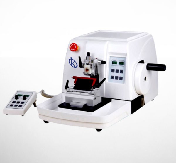
Rotating microtomes, also known as Minot, have a fixed blade and a mobile sample holder. The name of the rotation microtome is given because the sample holder is operated by a handwheel. The rotation movement of the steering wheel is transformed into a straight movement. Normally the sample holder of these microtomes moves in the downward direction. The prepared samples accumulate on the blade. The advantage of these microtomes is that the high flywheel mass matches the different hardnesses in the same test, resulting in a uniform cut. Rotational microtomes allow to prepare samples between 1 and 60 µm.
What are its advantages?
- By having more weight, it has more precision, allows to obtain very fine serial sections.
- The advance mechanism is more accurate.
What considerations should I take into account?
- The high price due to the complexity of the advancement mechanism, which also makes repairs more difficult and expensive.
- The inability to cut tissues included in celloidin, gelatin and propylene glycol with it.
In Kalstein we offer you sophisticated microtomes that will allow you to offer the best analysis since they have the best technology. That’s why we invite you to take a look at our available microtomes at HERE