An optical microscope is an instrument by which we can observe elements or structures that cannot be observed with the naked eye, through the use of lenses, viewfinders and light rays, which approach or enlarge the image at scales suitable for examination and subsequent study.
Microscope, like all laboratory equipment, has been evolving day by day thanks to technological advances that have brought a series of improvements and refinements of its optical, electronic and mechanical components. Therefore, it is of vital importance to know the basic structure of a microscope and the parts that constitute it, as well as its specific function, which will greatly help the user in its interrelation with the microscopic equipment and thus be able to make the appropriate decisions about the necessary care and uses of it.
A light microscope is an optical device used to amplify and resolve fine details of a microscopic object. It is made up of different components: mechanical, optical and lighting.
Mechanical components of a microscope
- Base or foot: it supports the microscope and is usually of sufficient weight to give stability to the equipment.
- Column: attaches the platinum to the base and holds the condenser and diaphragm.
- Tube: it holds the eye and the target and keeps them separated by the correct working distance.
- Arm: continuation of the base or foot that allows to mobilize the microscope with the hand.
- Platinum: hold the preparations with the sample placed on a central perforation that lets light coming from the condenser or mirror through.
- Tweezers: hold the slide firmly on the plate.
- Stir: alternatively, it allows to place in working position the objectives of the microscope.
- Macrometric screw: move the deck or tube up or down, quickly zooming in or out of the target to the approximate working distance from the sample.
- Micrometric screw: allows the tube to be moved slowly and precisely to the deck or vice versa.
Optical components of a microscope
- Ocular: composed of lenses multiplying the increase of the target. They are located at the top of the tube. A good eyepiece (10x) can allow a total increase of 1000x.
- Objectives: composed of lenses of different magnification. They are located at the bottom of the tube (revolver system). A light microscope commonly has three lens targets: 10X, 40X and 100X, independent and interchangeable. Each of them has a given magnification, and their purpose is to magnify the object to produce a real image that is projected to the focal plane of the ocular.
Illumination system of a microscope
- Light source: artificial light from an external lamp or a lamp inserted into the base or foot of the microscope may be used. A mirror is required when using the external lamp.
- Mirror: This is needed by microscopes working with external lamps and allows the light to be reflected upwards through the diaphragm, the platinum, the preparation and the optical system. The flat mirror is used when there is enough light and the concave when there is little light.
- Condenser: concentrate the light beam in preparation.
- Diaphragm: regulates the amount of light that will pass through the preparation.
What do we offer you in Kalstein?
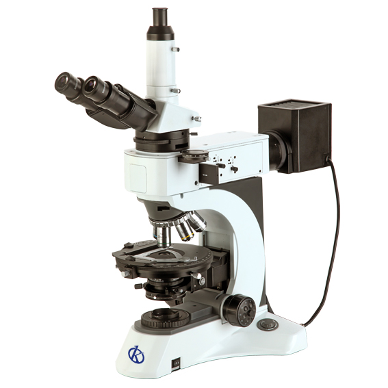
At Kalstein we are MANUFACTURERS of laboratory equipment of the highest quality and with the best technology. This time we present you our polarization microscope YR0262, which provides high quality images and contrast with a special tension-free lens. This equipment has the following characteristics that make it a novel microscope:
- Unique polarization, orthogonal polarization and conoscopic observation.
- Transmitted light and reflected light, 24V/100W long-lasting halogen lamp.
- Stage with 45° located, connecting mechanical stage with large range of motion, high efficiency without interference when converting targets.
For more information we invite you to take a look at: HERE