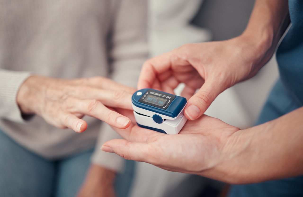
Pulse oximetry is a non-invasive method that allows the estimation of oxygen saturation of arterial hemoglobin and also monitors heart rate and pulse amplitude.
What is your story?
The first advances in the concept of oximetry were made in 1918 during the First World War when Krogh in Copenhagen tried to measure the oxygenation of pilots. In 1930 Millikan and Wood developed a two-wavelength pinnacle oximeter and in 1949 Wood and Geraci were able to measure the absolute oxygen saturation through photoelectric determination in the earlobe. In 1974, engineer Takuo Ayoagi of the Nihon Kohden, based on the fact that the arterial pulsations change the color of the blood and can be read using the radius of the absorption of red and infrared light, developed the first pulse oximeter.
In 1977 Minolta markets the “Oximet” by adding two optical fiber sensors. Subsequently, clinical trials are carried out at Stanford University and in 1981 “Biox and Nellcor” add the light sensors and the pulsatile signal that are currently used in clinical practice.
How does a pulse oximeter operate?
To determine the saturation of arterial hemoglobin with oxygen (SpO2), the pulse oximeter or pulse oximeter uses spectrophotometry based on which the oxyhemoglobin or oxygenated hemoglobin (HbO2) and the deoxyhemoglobin or reduced hemoglobin (Hb) absorb and transmit certain lengths of light spectrum wave for red light (640-660nm) and infrared light (910-940nm). HbO2 absorbs infrared light more and allows red light to pass through; on the contrary, Hb absorbs more red light (R) and allows infrared (IR) light to pass through. The radius of light absorption R and IR measures the degree of oxygenation of hemoglobin.
What is the difference between an oxygen measurement obtained with an oximeter and that obtained with an arterial gasometry?
Oximeters indirectly measure the amount of oxygen carried in the blood. Arterial gasometry directly measures both the amount of oxygen and the amount of gases (oxygen and carbon dioxide) that your blood contains. Arterial blood gas is obtained by drawing blood directly from the artery, which is painful. Oximetry is painless but not as accurate as arterial blood gas. In addition, the pulse oximeter does not measure carbon dioxide levels.
How factors can affect the accuracy of measurements in pulse oximeters?
The validity and reliability of conventional pulse oximeter measurements may be affected by various circumstances:
• The movement: this is the most common, especially in very young children or newborns. The key premise of conventional pulse oximetry was that the only pulsating component in motion was arterial blood.
• Low perfusion: the perfusion of the vascular bed between the light emitting diode (LED) and the sensor of the monitor probe determines the magnitude of the signal available for the pulse oximeter. By decreasing perfusion, so does the magnitude of the signal, as arterial pulsation is necessary for measurement, low perfusion states such as shock, low cardiac output and hypothermia can alter readings.
• Skin pigmentation and nail paint: dark skin could potentially have errors with SpO2 readings below 80% and nail polish, absorbs light at 660 nm or 940 nm can interfere with the ability of the pulse oximeter to interpret the SaO2.
• Electromagnetic interference: external electromagnetic energy such as that from tomographs, electrocautery, cell phones or others can cause interference of the correct reading of the oximeter and also produce an overheating of the sensor, which leads to low SpO2 readings and false alarms.
• Ambient light interference: intense white or red light can interfere with the reading of oximeters because they alter the function of photodetectors. This difficulty can be avoided by covering the sensor with a non-transparent material.
At Kalstein we offer you excellent and reliable pulse oximeters. That’s why we invite you to take a look at: HERE