It is a medical electronic equipment with a long history in which they have allowed a great advance in the field of medicine that captures the cardiac activity of the patient, which records any damage, signal, the existence of abnormal palpitations, size of the heart and position of their chambers, in addition to the effects caused by drugs or certain devices that control the heart (pacemaker).
The work of the electrocardiograph, as a clinical diagnostic equipment, is based on the installation of a series of electrodes on the surface of the patient’s skin at the level of the thoracic region and extremities. The result of this analysis is a document that records the data called electrocardiogram.
Through this study, you can monitor heart disease, such as: Irregular heartbeat (arrhythmia), blocked arteries, heart damage, heart failure, heart attack. These equipment, used in ambulances, emergency rooms and hospitals to diagnose a heart attack, is sometimes included in a routine examination of middle-aged or older adults because the risk of heart disease increases with age.
Parts of the electrocardiograph
Below we will see the parts of this instrument to perform electrocardiograms.
- Biopotentials amplifier: is composed of five parts, which are: Protection circuit, calibration signal, preamp, insulation circuit and amplifier driver.
- Right leg registration: it is formed by a ground that activated by the leg, controls the voltages that the patient receives and thus increase safety.
- Bypass Module: it is an amplified system of biopotentials that receives the signal of the electrodes by means of the resistances, achieving the derivation of interest.
- Signal Storage: It stores records of each patient’s data and stores the signal before printing via a digital keyboard.
- Microcontroller: controls each procedure performed by the electrocardiograph. The medical personnel in charge have several modes of operation with predetermined procedures. An example would be to be able to record twelve leads with four beats in each one or by fixed time segments.
- Registrar: by means of the signal sent by the electrodes, the equipment allows to give a printed result made with pens and continuous thermal paper.
How to use the electrocardiograph
By means of specific electronic signals received by this equipment, medical personnel can interpret the diagnosis in a less invasive way, which detects any cardiac abnormality. The detail is in obtaining the biosignals of the patient’s body through the main stages:
- To identify the physical variable to be measured by the study of the electrocardiogram allows measuring how ions influence cardiac cells to produce muscle contraction and relaxation, located in the patient’s chest.
- Sensor or translator, allows converting through chemical signals, the flow of ions in the heart muscle into an electrical current.
- Pre-amplification, increases the magnitude of the signal.
- Filtration, digitalization and transformation is the transformation of physical analog signals of the body and modern digital signals managed by computer systems, obtained and transformed from digital signals visualized in a disk, to be stored and sent.
For the use of this equipment, the risks are minimal, which the patient may feel anxiety, mild irritability in the skin caused by electrodes. There is no risk of electric shock. The electrocardiograph doesn’t send any electricity to the body. Just record the electricity.
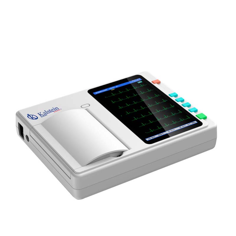
Electrocardiograph in KALSTEIN
At Kalstein we have the different teams with the best medical technology on the market. In addition, we have the best electrocardiographs, such as the 12-channel ECG machine, which allows to collect 12-lead ECG signals simultaneously and print ECG waveforms with a thermal printing system. The electrocardiogram is commonly used to detect abnormal heart rhythms and to investigate the cause of chest pains. An electrocardiogram records the electrical activity of the heart. The heart produces tiny electrical impulses that spread through the heart muscle to cause the heart to contract. These impulses can be detected by the ECG machine.
We offer equipment with model to YR, 7″ touch screen, recording mode thermal printing system. For a better specification, visit us HERE
We are manufacturers and we have the best advice to make your purchase the safest and at excellent prices, the best in the market. Follow us at HERE