At some point you’ve probably wondered how an electrocardiograph works? This medical device is part of a science called bioinstrumentation, a branch of biomedical engineering. It is responsible for recovering specific biosignals from the human body and then processing them in such a way that doctors can interpret them to obtain a diagnosis of the patient in the least invasive way possible.
An electrocardiograph is a clinical diagnostic medical team, which is responsible for capturing and expanding the electrical activity of the patient’s heart through the use of electrodes. The recording of this activity is called an electrocardiogram (ECG), which is defined as the continuous recording of electrical impulses in the heart, and is commonly used by doctors to determine any damage to the heart, if abnormal heart palpitations, the size and position of the heart chambers, and effects of drugs or devices used to control the heart (such as pacemakers) exist.
What is the basis of the operation of an electrocardiograph?
The function of the electrocardiograph is based on the placement of a series of electrodes on the surface of the patient’s skin at the level of the chest region and extremities. The electrode on the skin is connected to another electrode through the electrocardiograph. A galvanometer measures the current passing through the apparatus and is transmitted directly to the inscriptor to record the waves and complexes that reflect the electrical activity of the heart.
In order to correctly pick up the electrical signal from the heart it is necessary to place the electrodes in 6 precordial positions and in the same way in the four extremities. The way these electrodes are arranged on the body surface determines different electrical configurations, supplying different electrocardiographic leads. When the equipment captures the electrical spectrum of the heart, it inscribes in the millimeter paper the spectrum recorded by the electrodes.
Parts of an electrocardiograph
- Bioppontential amplifier: It consists of the protection circuit, calibration signal, preamplifier, insulation circuit and amplifier manager.
- Right leg circuit: This part of the electrocardiograph has the ability to generate active earth isolated from the electrical earth of the circuit in the patient’s right leg in order to reduce the voltages it receives, and consequently increase its safety. This is achieved by reducing the impedance of the ground electrode.
- Bypass selector: It consists of a module that is coupled to a biopotentials amplification system. The module ponders the contribution of each electrode through resistors, achieving the derivation of interest.
- Memory System: It is a memory where the signal is stored before being printed along with the data entered manually via a digital keyboard. A digital analog converter is used to convert the signal.
- Microcontroller: This device dominates every procedure performed by the electrocardiograph. The operator can select different operation modes with pre-programmed processes.
- Registrar: It is the device responsible for printing the signal that the electrodes pick up, that is, the result of the electrocardiogram. It uses pens and continuous thermal paper or inkjet.
How to interpret the results of an electrocardiograph?
In order to interpret the results emitted by the electrocardiograph, the first thing you need to know is which waves you will see reflected in the ECG. These waves are identified with letters of the alphabet from the letter P to the letter L. Similarly, to know whether or not a result is within the normal parameters or not, you need to know which wave has to be positive or negative. In addition to knowing the size of them, in amplitude and length, because any alteration in them will be reflected in the ECG.
To find out if the wave is OK, you must count the ECG frames and these must be within normal parameters. Reading an ECG isn’t easy. It’s actually the doctors’ job. However, for a normal person it would not be impossible to learn to read and interpret an electrocardiogram. The ECG provides very important information about the electrical activity of the heart, so its results allow you to quickly take action and prevent any heart problems or mishaps.
What do we offer you in Kalstein?
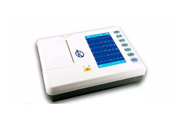
Kalstein is a company MANUFACTURER of medical equipment and laboratory of the highest quality and technology, with the best prices in the market, so you can make your purchase confidently with us, knowing that you have the service and advice of a solid company and with a lot of experience in the field. In this opportunity we present our Electrocardiograph YR05161, This new 6 channel YR ECG gives you the power of advanced and easy to use technologies, multiple forms of data management, as well as accurate measurements and interpretations, which can improve the diagnostic confidence of cardiologists.
For more information we invite you to take a look: HERE