The microtome is a mechanical instrument used to cut biological samples into very thin segments for microscopic examination. Biological samples can be embedded and presented in many ways for sectioning. But more often, these samples are embedded in paraffin wax blocks and the most common way to section these samples is by microtome.
The older form of the microtome allowed manual cutting of fresh or fixed material with a sharp razor. Modern microtomes are precision instruments designed to cut thin sections evenly from a variety of materials for detailed microscopic examination. A fundamental element for the production of good sections is the microtome knife.
Microtomy virtually begins and ends with a sharp, flawless cutting edge. The introduction of disposable blades has facilitated the production of good quality thin sections, but they are often not satisfactory for sectioning harder tissues, especially bone. A sharp, blemish-free knife edge is essential for producing good sections. Since many types of microtome are commercially available on the market, choosing the correct microtome is essential to produce the best result as needed.
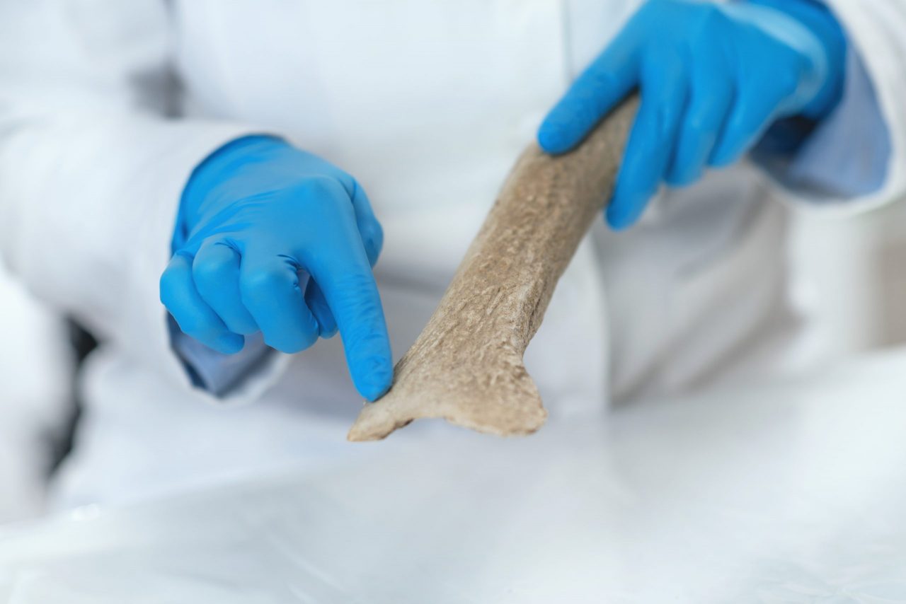
How to cut bones with a microtome?
The value of the histological examination in the diagnosis and classification of clinical conditions is entrusted to the experience of the pathological anatomy laboratory in handling the wide range of specimen types submitted for analysis.
From receipt of the tissue sample to presentation of the slide for microscopic examination, histologists must consider the composition of the specimen to determine how it should be handled effectively.
Most samples follow a routine cycle of dehydration and wax embedding with paraffin for the purpose of sectioning on the microtome. However, due to its high calcium content, bone is a particularly difficult tissue to section and the density of the sample must be taken into consideration. Cutting decalcified sections of bone requires embedding in a resin, a specialized microtome, and modified staining techniques.
Consequently, most laboratories decalcify bone samples, allowing them to be embedded in paraffin and processed. Nitric acid and formic acid are used as decalcification agents, with formic acid being more suitable for laboratories performing molecular analysis, because unlike nitric acid, it does not destroy DNA.
Descaling varies in time depending on the size and type of bone sample. The quality of a section can also have a major impact on the accuracy and reliability of the diagnosis, and is related to the selection of the most appropriate microtome.
What is recommended?
On many occasions, manual sectioning is recommended for paraffin-embedded specimens due to the need to vary the levels of force used to cut various parts of the sample.
Tissues common to analysis, on the other hand, sometimes require larger and thicker sections. As a consequence, these samples sometimes require casting in resin with the use of a fully mechanized microtome that slowly applies force in a constant and controlled manner to thereby generate sections of good quality.
At Kalstein we offer you a wide range of innovative microtomes and pathological anatomy equipment such as specialized bath for histology, paraffin dispensers, cooling plates, tissue processing machines, paraffin inclusion stations. That is why we invite you to take a look at the “Products” HERE